News

Publication Placeholder

Santarelli, S., Zimmermann, C., Kalideris, G., Lesuis, S.L., Arloth, J., Uribe, A., Dournes, C., Balsevich, G., Hartmann, J., Masana, M., Binder, E.B., Spengler, D., and Schmidt, M.V.
Psychoneuroendocrinology, 2017,  78, 213-221.

An adverse early life environment can enhance stress resilience in adulthood.

Chronic stress is a major risk factor for depression. Interestingly, not all individuals develop psychopathology after chronic stress exposure. In contrast to the prevailing view that stress effects are cumulative and increase stress vulnerability throughout life, the match/mismatch hypothesis of psychiatric disorders. The match/mismatch hypothesis proposes that individuals who experience moderate levels of early life psychosocial stress can acquire resilience to renewed stress exposure later in life. Here, we have tested this hypothesis by comparing the developmental effects of 2 opposite early life conditions, when followed by 2 opposite adult environments. Male Balb/c mice were exposed to either adverse early life conditions (limited nesting and bedding material) or a supportive rearing environment (early handling). At adulthood, the animals of each group were either housed with an ovariectomized female (supportive environment) or underwent chronic social defeat stress (socially adverse environment) for 3 weeks. At the end of the adult manipulations, all of the animals were returned to standard housing conditions. Then, we compared the neuroendocrine, behavioral and molecular effects of the interaction between early and adult environment. Our study shows that early life adversity does not necessarily result in increased vulnerability to stress. Specific endophenotypes, like hypothalamic-pituitary-adrenal axis activity, anxiety-related behavior and glucocorticoid receptor expression levels in the hippocampus were not significantly altered when adversity is experienced during early life and in adulthood, and are mainly affected by either early life or adult life adversity alone. Overall our data support the notion that being raised in a stressful environment prepares the offspring to better cope with a challenging adult environment and emphasize the role of early life experiences in shaping adult responsiveness to stress.

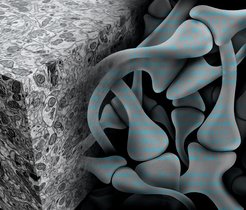
 

How does consciousness arise? Researchers suspect that the answer to this question lies in the connections between neurons. Unfortunately, however, little is known about the wiring of the brain. This is due also to a problem of time: tracking down connections in collected data would require man-hours amounting to many lifetimes, as no computer has been able to identify the neural cell contacts reliably enough up to now. Scientists from the Max Planck Institute of Neurobiology in Martinsried plan to change this with the help of artificial intelligence. They have trained several artificial neural networks and thereby enabled the vastly accelerated reconstruction of neural circuits.

Neurons need company. Individually, these cells can achieve little, however when they join forces neurons form a powerful network which controls our behaviour, among other things. As part of this process, the cells exchange information via their contact points, the synapses. Information about which neurons are connected to each other when and where is crucial to our understanding of basic brain functions and superordinate processes like learning, memory, consciousness and disorders of the nervous system. Researchers suspect that the key to all of this lies in the wiring of the approximately 100 billion cells in the human brain.

To be able to use this key, the connectome, that is every single neuron in the brain with its thousands of contacts and partner cells, must be mapped. Only a few years ago, the prospect of achieving this seemed unattainable. However, the scientists in the Electrons – Photons – Neurons Department of the Max Planck Institute of Neurobiology refuse to be deterred by the notion that something seems “unattainable”. Hence, over the past few years, they have developed and improved staining and microscopy methods which can be used to transform brain tissue samples into high-resolution, three-dimensional electron microscope images. Their latest microscope, which is being used by the Department as a prototype, scans the surface of a sample with 91 electron beams in parallel before exposing the next sample level. Compared to the previous model, this increases the data acquisition rate by a factor of over 50. As a result an entire mouse brain could be mapped in just a few years rather than decades. 

Although it is now possible to decompose a piece of brain tissue into billions of pixels, the analysis of these electron microscope images takes many years. This is due to the fact that the standard computer algorithms are often too inaccurate to reliably trace the neurons’ wafer-thin projections over long distances and to identify the synapses. For this reason, people still have to spend hours in front of computer screens identifying the synapses in the piles of images generated by the electron microscope.

However the Max Planck scientists led by Jörgen Kornfeld have now overcome this obstacle with the help of artificial neural networks. These algorithms can learn from examples and experience and make generalizations based on this knowledge

read more


Publication Placeholder

Sherrard, R., Luehr, S., Holzkamp, H., McJunkin, K., Memar, N., and Conradt, B.
Genes Dev, 2017, 31, 209-222.

miRNAs cooperate in apoptosis regulation during C. elegans development.

Programmed cell death occurs in a highly reproducible manner during Caenorhabditis elegans development. We demonstrate that, during embryogenesis, miR-35 and miR-58 bantam family microRNAs (miRNAs) cooperate to prevent the precocious death of mothers of cells programmed to die by repressing the gene egl-1, which encodes a proapoptotic BH3-only protein. In addition, we present evidence that repression of egl-1 is dependent on binding sites for miR-35 and miR-58 family miRNAs within the egl-1 3' untranslated region (UTR), which affect both mRNA copy number and translation. Furthermore, using single-molecule RNA fluorescent in situ hybridization (smRNA FISH), we show that egl-1 is transcribed in the mother of a cell programmed to die and that miR-35 and miR-58 family miRNAs prevent this mother from dying by keeping the copy number of egl-1 mRNA below a critical threshold. Finally, miR-35 and miR-58 family miRNAs can also dampen the transcriptional boost of egl-1 that occurs specifically in a daughter cell that is programmed to die. We propose that miRNAs compensate for lineage-specific differences in egl-1 transcriptional activation, thus ensuring that EGL-1 activity reaches the threshold necessary to trigger death only in daughter cells that are programmed to die.


Publication Placeholder

Kremer, M.C., Jung, C., Batelli, S., Rubin, G.M., and Gaul, U..
Glia, 2017, [Epub ahead of print].

The glia of the adult Drosophila nervous system.

Glia play crucial roles in the development and homeostasis of the nervous system. While the GLIA in the Drosophila embryo have been well characterized, their study in the adult nervous system has been limited. Here, we present a detailed description of the glia in the adult nervous system, based on the analysis of some 500 glial drivers we identified within a collection of synthetic GAL4 lines. We find that glia make up ∼10% of the cells in the nervous system and envelop all compartments of neurons (soma, dendrites, axons) as well as the nervous system as a whole. Our morphological analysis suggests a set of simple rules governing the morphogenesis of glia and their interactions with other cells. All glial subtypes minimize contact with their glial neighbors but maximize their contact with neurons and adapt their macromorphology and micromorphology to the neuronal entities they envelop. Finally, glial cells show no obvious spatial organization or registration with neuronal entities. Our detailed description of all glial subtypes and their regional specializations, together with the powerful genetic toolkit we provide, will facilitate the functional analysis of glia in the mature nervous system. GLIA 2017.


Publication Placeholder

Matscheko, N., Mayrhofer, P., and Wollert, T.
Autophagy, 2017, [Epub ahead of print].

Passing membranes to autophagy: Unconventional membrane tethering by Atg17.

Macroautophagy delivers cytoplasmic material to lysosomal/vacuolar compartments for degradation. Conserved multi-subunit complexes, composed of autophagy related (Atg) proteins, initiate the formation of membrane precursors, termed phagophores. Under physiological conditions these cup- shaped structures can capture cytoplasmic material highly selectively. Starvation or cytotoxic stresses, however, initiate the formation of much larger phagophores to enclose cytoplasm nonselectively. The biogenesis of nonselective autophagosomes is initiated by the hierarchical assembly of the Atg1 kinase complex and the recruitment of Atg9 vesicles at the phagophore assembly site (PAS). In this punctum we summarize our recent findings regarding tethering of Atg9 vesicles by the Atg1 kinase complex. We discuss membrane tethering by and activation of its central subunit Atg17 in the context of other canonical membrane tethering factors. Our results show that Atg17 suffices to bind and tether Atg9 vesicles. The Atg31-Atg29 subcomplex inhibits Atg17 activity, and activation of Atg17 depends on the formation of the Atg1 kinase complex that involves recruiting Atg1-Atg13. Our studies lead to a model of unconventional membrane tethering in autophagy.


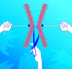
Once together, never apart – isn’t that how the saying goes? Not so in meiosis, the special type of cell division in which gametes, sperm and egg cells are formed. At the start of meiosis the ring-shaped protein complex, referred to as cohesin, is the string that ties the chromosome strands together. The chromosomes are where the blueprint for the body is stored. If each egg cell and each sperm is to come out of meiosis with only one set of chromosomes, these strings need to be cut up in a precise pattern. Scientists from the Max Planck Institute of Biochemistry have demonstrated in baker’s yeast how a kinase enzyme, which is also present in the human body, controls the cleavage of the cohesin rings and coordinates it with the exit from meiosis and gamete formation. It is a mechanism, which could explain how chromosome segregation is regulated, or goes wrong, in human sperm and egg cells.

Why is it that children look like their parents? Most of the cells in our body are diploid, which means that they have two copies of each chromosome – one from the mother and one from the father. Only gametes, in other words egg and sperm cells, contain a single copy. So the cell has to halve its double set of chromosomes in order to be able to make haploid gametes. This happens in a special type of cell division called meiosis. However, meiosis is surprisingly complicated. Instead of just dividing up the maternal and paternal chromosomes between two daughter cells, the chromosomes are first duplicated so that they each consist of two strands, or chromatids. Duplicated paternal and maternal chromosomes then align and the chromosome arms are spliced together. This produces new chromosomes out of four chromatids held together by ring-shaped protein complexes, the cohesins, which act like strings. Two divisions of the nucleus are needed in order to split these chromosomes up into their chromatids again in processes referred to as the first and the second meiotic divisions. In the first division, the cohesin rings on the chromosome arms are cut up by the enzyme separase, which acts like a molecular pair of scissors. This results in X-shaped chromosomes consisting of two chromatids held together by cohesin rings only in their centre, known as the centromer. The second meiotic division is when the separase scissors ultimately cut the cohesin rings at the centromer. Four haploid gamete nuclei are produced, each of which contains one chromatid from each chromosome, in other words a blueprint for the body. If the egg is fertilized, the genetic material from the mother and the father combines and a new diploid chromosome set is produced, or to put it simply: a baby with daddy’s nose and mummy’s eyes.

read more


Publication Placeholder

Princz, L.N., Wild, P., Bittmann, J., Aguado, F.J., Blanco, M.G., Matos, J., and Pfander, B.
EMBO J, 2017, [Epub ahead of print].

Dbf4-dependent kinase and the Rtt107 scaffold promote Mus81-Mms4 resolvase activation during mitosis.

DNA repair by homologous recombination is under stringent cell cycle control. This includes the last step of the reaction, disentanglement of DNA joint molecules (JMs). Previous work has established that JM resolving nucleases are activated specifically at the onset of mitosis. In case of budding yeast Mus81-Mms4, this cell cycle stage-specific activation is known to depend on phosphorylation by CDK and Cdc5 kinases. Here, we show that a third cell cycle kinase, Cdc7-Dbf4 (DDK), targets Mus81-Mms4 in conjunction with Cdc5-both kinases bind to as well as phosphorylate Mus81-Mms4 in an interdependent manner. Moreover, DDK-mediated phosphorylation of Mms4 is strictly required for Mus81 activation in mitosis, establishing DDK as a novel regulator of homologous recombination. The scaffold protein Rtt107, which binds the Mus81-Mms4 complex, interacts with Cdc7 and thereby targets DDK and Cdc5 to the complex enabling full Mus81 activation. Therefore, Mus81 activation in mitosis involves at least three cell cycle kinases, CDK, Cdc5 and DDK Furthermore, tethering of the kinases in a stable complex with Mus81 is critical for efficient JM resolution.


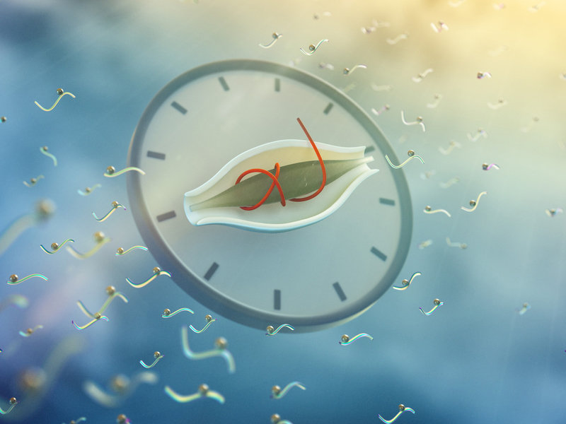
Circadian is the latin meaning for “about a day”. Circadian clocks have evolved to adapt our lives to the daily environmental changes on earth: light and warmth during the day and darkness and cold at night. Scientists at the Max-Planck-Institute of Biochemistry in Martinsried discovered with the help of the mass spectrometry, that more than 25 percent of the molecular protein switches in mouse liver cells change in a daily manner. These rhythmic switches are binding sites for phosphate molecules, that regulate the function of proteins, and thereby the daily metabolic processes in the organ. The study was published in the journal Cell Metabolism.

read more


Publication Placeholder

Bantele, S.C., Ferreira, P., Gritenaite, D., Boos, D., and Pfander, B.
Elife, 2017, 6.

Targeting of the Fun30 nucleosome remodeller by the Dpb11 scaffold facilitates cell cycle-regulated DNA end resection.

DNA double strand breaks (DSBs) can be repaired by either recombination-based or direct ligation-based mechanisms. Pathway choice is made at the level of DNA end resection, a nucleolytic processing step, which primes DSBs for repair by recombination. Resection is thus under cell cycle control, but additionally regulated by chromatin and nucleosome remodellers. Here we show that both layers of control converge in the regulation of resection by the evolutionarily conserved Fun30/SMARCAD1 remodeller. Yeast Fun30 and human SMARCAD1 are cell cycle-regulated by interaction with the DSB-localized scaffold proteins Dpb11 and TOPBP1, respectively. In yeast this protein assembly additionally comprises the 9-1-1 damage sensor, is involved in localizing Fun30 to damaged chromatin and thus is required for efficient long-range resection of DSBs. Notably, artificial targeting of Fun30 to DSBs is sufficient to bypass the cell cycle regulation of long-range resection, indicating that chromatin remodelling during resection is underlying DSB repair pathway choice.


Publication Placeholder

Thanisch, K., Song, C., Engelkamp, D., Koch, J., Wang, A., Hallberg, E., Foisner, R., Leonhardt, H., Stewart, C.L., Joffe, B., and Solovei, I.
Differentiation, 2017, 94, 58-70.

Nuclear envelope localization of LEMD2 is developmentally dynamic and lamin A/C dependent yet insufficient for heterochromatin tethering.

Peripheral heterochromatin in mammalian nuclei is tethered to the nuclear envelope by at least two mechanisms here referred to as the A- and B-tethers. The A-tether includes lamins A/C and additional unknown components presumably INM protein(s) interacting with both lamins A/C and chromatin. The B-tether includes the inner nuclear membrane (INM) protein Lamin B-receptor, which binds B-type lamins and chromatin. Generally, at least one of the tethers is always present in the nuclear envelope of mammalian cells. Deletion of both causes the loss of peripheral heterochromatin and consequently inversion of the entire nuclear architecture, with this occurring naturally in rod photoreceptors of nocturnal mammals. The tethers are differentially utilized during development, regulate gene expression in opposite manners, and play an important role during cell differentiation. Here we aimed to identify the unknown chromatin binding component(s) of the A-tether. We analyzed 10 mouse tissues by immunostaining with antibodies against 7 INM proteins and found that every cell type has specific, although differentially and developmentally regulated, sets of these proteins. In particular, we found that INM protein LEMD2 is concomitantly expressed with A-type lamins in various cell types but is lacking in inverted nuclei of rod cells. Truncation or deletion of Lmna resulted in the downregulation and mislocalization of LEMD2, suggesting that the two proteins interact and pointing at LEMD2 as a potential chromatin binding mediator of the A-tether. Using nuclei of mouse rods as an experimental model lacking peripheral heterochromatin, we expressed a LEMD2 transgene alone or in combination with lamin C in these cells and observed no restoration of peripheral heterochromatin in either case. We conclude that in contrary to the B-tether, the A-tether has a more intricate composition and consists of multiple components that presumably vary, at differing degrees of redundancy, between cell types and differentiation stages.