News

Publication Placeholder

Schopf FH, Biebl MM, Buchner J.
Nat Rev Mol Cell Biol, 2017, [Epub ahead of print].

The HSP90 chaperone machinery.

The heat shock protein 90 (HSP90) chaperone machinery is a key regulator of proteostasis under both physiological and stress conditions in eukaryotic cells. As HSP90 has several hundred protein substrates (or 'clients'), it is involved in many cellular processes beyond protein folding, which include DNA repair, development, the immune response and neurodegenerative disease. A large number of co-chaperones interact with HSP90 and regulate the ATPase-associated conformational changes of the HSP90 dimer that occur during the processing of clients. Recent progress has allowed the interactions of clients with HSP90 and its co-chaperones to be defined. Owing to the importance of HSP90 in the regulation of many cellular proteins, it has become a promising drug target for the treatment of several diseases, which include cancer and diseases associated with protein misfolding.


 

Publication Placeholder

Zhang X, Schnorrer F.
FEBS J. 2017, 284, 1178-1181.

AIDing-targeted protein degradation in Drosophila.

Conditional protein depletion is highly desirable for investigating protein functions in complex organisms. In this issue, Bence and colleagues combined auxin-inducible degradation with CRISPR, establishing an elegant tool to control protein levels. They achieve precise spatio-temporal control of protein degradation during Drosophila oogenesis and early embryogenesis by combining suitable GAL4 drivers (spatial control) with auxin feeding protocols (temporal control).


Biochemist Karl-Peter Hopfner studies how cells detect and repair the damage to their DNA. His work has now won him Germany’s most prestigious prize for research.

The magnitude of the repair job is mind-boggling: Every day, around 100,000 of the building-blocks (‘bases’) in the genomic DNA of virtually every cell in our bodies suffer damage, and are chemically altered. Left unrepaired, these alterations in our genetic material can kill cells, induce the development of tumors, precipitate premature ageing and cause congenital diseases when they occur in germ cells. However, evolution has equipped cells with highly efficient mechanisms for the repair of DNA damage.

At LMU’s Gene Center Karl-Peter Hopfner is studying the complex molecular machines that make it possible for cells to locate adventitious damage and enzymatic errors and repair or remove the modified bases. With the aid of high-resolution methodologies such as X-ray crystallography and cryo-electron microscopy, Hopfner elucidates the structures and modes of action of the molecular machines that tackle this gargantuan task. In order to develop effective treatments for disorders that result when these systems themselves are defective, detailed knowledge of their molecular form is an essential prerequisite. The Deutsche Forschungsgemeinschaft (DFG) has now acknowledged Karl-Peter Hopfner’s contributions to research by awarding him a Leibniz Prize.

read more


Borst 2017

In order to react to changes in the environment in good time, the brain must analyze the signals it receives from the eyes rapidly and accurately. For example, the ability to recognise the direction in which an approaching car is moving is vital to the survival of modern humans in cities. Using the brain of the fruit fly Drosophila as a model, scientists from the Max Planck Institute of Neurobiology study how the brain extracts this essential motion information. They have now described in detail the cells that enable downstream neurons to recognize the direction of movement. Interestingly, the characteristics of these input cells exactly match to a motion detector model they recently proposed. In addition, the cells alter their characteristics according to the animals’ state: when the fly is active, the cells respond faster to light stimuli.

Humans perceive their environment mainly through their eyes. The ability to recognize movements and their direction is something that seems almost trivial and automatic to us. However, this information has to be processed in the brain as the light-sensitive sensory cells of the retina can only register changes in contrast. The direction of a movement can only be calculated through the comparison of neighbouring signals. Various models exist for these calculations. Alexander Borst and his team at the Max Planck Institute of Neurobiology study the extent to which these models can be applied to the brain’s neuronal circuitry. Their test subject is the fruit fly Drosophila, a master of motion perception.

read more


Although all cells in an organism contain the same genes, only some of the genes are activated in a given cells and others remain inactive. Genes coil around histone proteins in the form of DNA threads. If a gene has to remain inactive, its histones are marked by the PRC2 enzyme so that this gene is locked down and cannot be read. When cells divide and the genes are copied, these histone marks must be placed again, at exactly the same location. The mechanism that enables transmission of this information has now been explained by Jürg Müller from the Max Planck Institute of Biochemistry in Martinsried in a study published in the journal Science.

In animals and plants, the genomic DNA in the cell nucleus is wrapped around small proteins known as histones. Jürg Müller, Leader of the Biology of Chromatin Research Group at the MPI of Biochemistry explains: “The DNA is like a big library of books. Each book contains the instruction manual for making a protein. Although the same DNA library is present in all cells, some of the books are ‘sealed’, so they cannot be read. A muscle cell requires other protein-building instructions than an intestinal cell.” An essential mechanism to prevent the expression of genes relies on the chemical marking of histone proteins to permanently “lock down” genes. In the current study, Müller and his team examined how such gene locks are transmitted during cell division. Histones play a key role in determining how accessible a gene is. When genes need to be permanently locked down, their histones are chemically modified by the enzyme PRC2. “If we imagine the histones as the binder of the book, PRC2 helps to seal that book and prevent that it gets opened and read,” explains Müller. 

read more


Publication Placeholder

Laprell F, Finkl K, Müller J.
Science, 2017, [Epub ahead of print].

Propagation of Polycomb-repressed chromatin requires sequence-specific recruitment to DNA.

Epigenetic inheritance models posit that during Polycomb repression, Polycomb Repressive Complex 2 (PRC2) propagates histone H3K27 tri-methylation (H3K27me3) independently of DNA sequence. We show that insertion of Polycomb Response Element (PRE) DNA into the Drosophila genome creates extended domains of H3K27me3-modified nucleosomes in the flanking chromatin and causes repression of a linked reporter gene. After excision of PRE DNA, H3K27me3 nucleosomes become diluted with each round of DNA replication and reporter gene repression is lost, whereas in replication-stalled cells, H3K27me3 levels stay high and repression persists. Hence, H3K27me3-marked nucleosomes provide a memory of repression that is transmitted in a sequence-independent manner to daughter strand DNA during replication. In contrast, propagation of H3K27 tri-methylation to newly incorporated nucleosomes requires sequence-specific targeting of PRC2 to PRE DNA.


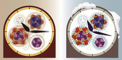
A team of Dutch and German researchers under the leadership of Albert Heck and Friedrich Förster has discovered the operation of one of the oldest biological clocks in the world, which is crucial for life on earth as we know it. The researcher from the Max Planck Institute of Biochemistry and the Utrecht University applied a new combination of cutting-edge research techniques. They discovered how the biological clock in cyanobacteria works in detail. Important to understand life, because cyanobacteria were the first organisms on earth producing oxygen via photosynthesis. The results of their research were published in Science.

Ten years ago, researchers discovered that the biological clock in cyanobacteria consists of only three protein components: KaiA, KaiB and KaiC. These are the building blocks - the gears, springs and balances - of an ingenious system resembling a precision Swiss timepiece. In 2005, Japanese scientists published an article in Science showing that a solution of these three components in a test tube could run a 24-hour cycle for days when a bit of energy was added. However, the scientists were not able to uncover the exact operation of the system, despite its relative simplicity.

William Faulkner
How could the scientists resolve the working of the individual pieces? “In the end, the trick to understand the ticking biological clock in cyanobacteria was to literally make time stop”, tells research leader Albert Heck from Utrecht University. “Or as William Faulkner, Nobel Prize Laureate in Literature said: ‘Only when the clock stops does time come to life.’ Faulkner spoke taking a pause in the constant haste of life. That was also the trick here. We slowed the biological clock by running it in the fridge for a week. In the literal sense we have frozen the time.”

read more


Publication Placeholder

Rieckmann, J.C., Geiger, R., Hornburg, D., Wolf, T., Kveler, K., Jarrossay, D., Sallusto, F., Shen-Orr, S.S., Lanzavecchia, A., Mann, M., and Meissner, F.
Nat Immunol, 2017, [Epub ahead of print].

Social network architecture of human immune cells unveiled by quantitative proteomics.

The immune system is unique in its dynamic interplay between numerous cell types. However, a system-wide view of how immune cells communicate to protect against disease has not yet been established. We applied high-resolution mass-spectrometry-based proteomics to characterize 28 primary human hematopoietic cell populations in steady and activated states at a depth of >10,000 proteins in total. Protein copy numbers revealed a specialization of immune cells for ligand and receptor expression, thereby connecting distinct immune functions. By integrating total and secreted proteomes, we discovered fundamental intercellular communication structures and previously unknown connections between cell types. Our publicly accessible (http://www.immprot.org/) proteomic resource provides a framework for the orchestration of cellular interplay and a reference for altered communication associated with pathology.


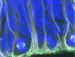
Neurodegenerative diseases such as Alzheimer‘s or Parkinson‘s, but also strokes or other types of traumatic brain damage, result in the death of nerve cells in the brain. Since the mammalian brain is capable of replacing nerve cells only in certain restricted regions, such nerve-cell loss is in most cases permanent. Similarly, the capacity to form new nerve cells in the mature brain is limited to specific areas. The cells responsible for neurogenesis in the mature brain are called adult neural stem cells, but little is known about their developmental origins. Now an international research collaboration led by Magdalena Götz, Professor of Physiological Genomics at LMU’s Biomedical Center and Director of the Institute for Stem Cell Research at the Helmholtz Zentrum Munich, has demonstrated that the mode of division of stem cells has a profound influence on the numbers of adult neural stem cells formed during embryonic development. The new findings appear in the journal Neuron.

read more


Publication Placeholder

Balaji, R., Bielmeier, C., Harz, H., Bates, J., Stadler, C., Hildebrand, A., and Classen, A.K.
Sci Rep, 2017, 7, 42786.

Calcium spikes, waves and oscillations in a large, patterned epithelial tissue.

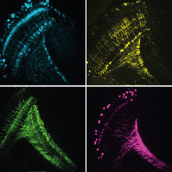
While calcium signaling in excitable cells, such as muscle or neurons, is extensively characterized, calcium signaling in epithelial tissues is little understood. Specifically, the range of intercellular calcium signaling patterns elicited by tightly coupled epithelial cells and their function in the regulation of epithelial characteristics are little explored. We found that in Drosophila imaginal discs, a widely studied epithelial model organ, complex spatiotemporal calcium dynamics occur. We describe patterns that include intercellular waves traversing large tissue domains in striking oscillatory patterns as well as spikes confined to local domains of neighboring cells. The spatiotemporal characteristics of intercellular waves and oscillations arise as emergent properties of calcium mobilization within a sheet of gap-junction coupled cells and are influenced by cell size and environmental history. While the in vivo function of spikes, waves and oscillations requires further characterization, our genetic experiments suggest that core calcium signaling components guide actomyosin organization. Our study thus suggests a possible role for calcium signaling in epithelia but importantly, introduces a model epithelium enabling the dissection of cellular mechanisms supporting the initiation, transmission and regeneration of long-range intercellular calcium waves and the emergence of oscillations in a highly coupled multicellular sheet.